Fundus Photography
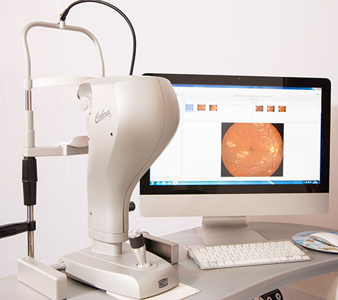
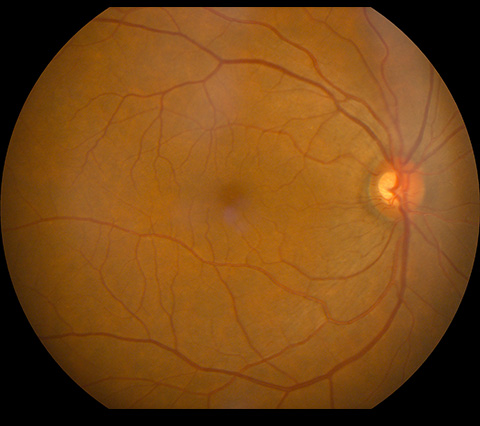
With its ergonomic design, it provides a detailed view of the entire fundus image with a true 45° field of view. This system offers images of the retina with minimal exposure to inflammation, which allows us to rapidly acquire a detailed view of the lower part of the eye, while minimizing patient discomfort. In addition, it uses a high-resolution CCD sensor (2 megapixels) for patient alignment (with IR illumination) and to capture retinal images with a flash of white light.